
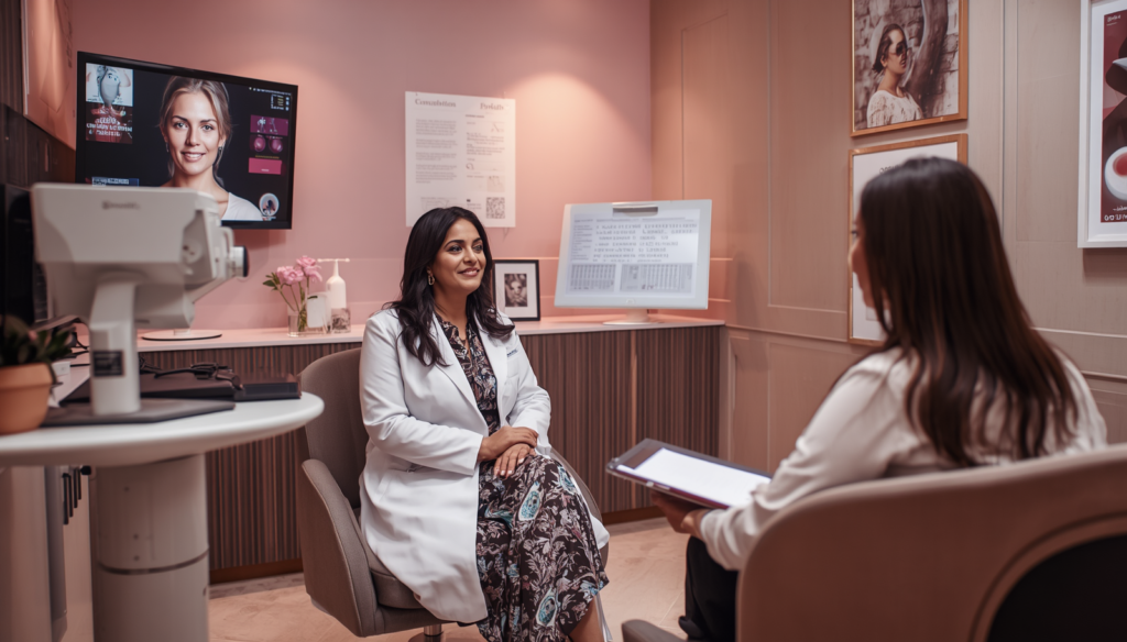
Decoding Pap Smear Terminology and Biopsy Findings
Receiving a notification that your routine cervical screening or Pap smear returned an “abnormal” result can naturally cause anxiety and uncertainty. However, an abnormal screening result is rarely an indicator of invasive cervical cancer. In over 90% of clinical cases, abnormal cytology simply highlights early, highly treatable cellular changes on the surface of the cervix—frequently driven by persistent, high-risk strains of the Human Papillomavirus (HPV).
Consulting an internationally accredited specialist recognized as a leading colposcopy specialist in dubai, such as Dr. Sunita Arora (MRCOG UK, MRCPI Ireland), provides women access to state-of-the-art diagnostic evaluation, optical magnification, and gentle, fertility-preserving treatment protocols designed to resolve precancerous lesions long before they pose a serious threat to health.

The Cervical Cytology & Histology Matrix: Understanding Pap Smear Results
A Pap smear acts as a broad primary screening tool that collects superficial epithelial cells from the ectocervix and endocervical canal. When these cells demonstrate structural atypia under microscopic examination, a colposcopy is the gold-standard diagnostic step used to evaluate the true depth and extent of tissue changes (histology).
By combining high-magnification colposcopic optics with selective cervical tissue sampling, your specialist can differentiate between temporary benign inflammation and true precancerous lesions known as Cervical Intraepithelial Neoplasia (CIN).
“Early identification of high-risk HPV strains through combined co-testing and colposcopic mapping allows us to intervene during the precancerous window. Conservative treatment of CIN 2 and CIN 3 lesions via precision loop excision effectively prevents over 95% of cervical cancers while fully preserving future reproductive capacity.”
American Board of Cosmetic Gynecology (ABCG)
Detailed Clinical Workflows: What to Expect During a Colposcopy
Undergoing a colposcopic examination is a straightforward, outpatient procedure performed in the comfort of a private clinic setting. The procedure requires no general anesthesia, takes approximately 15 to 20 minutes, and focuses on complete diagnostic precision.
1. High-Magnification Optical Inspection & Dynamic Staining
The patient is comfortably positioned, and a gentle speculum is placed to visualize the cervix. The colposcopy specialist in Dubai positions the colposcope—a high-powered binocular microscope with specialized green-filter light—a few inches outside the vagina. The cervix is carefully swabbed with a mild 3%–5% acetic acid (vinegar) solution and Lugol’s iodine.
2. Identification of Acetowhite Lesions & Vascular Patterns
Dysplastic cells containing higher concentrations of nuclear protein react to acetic acid by turning white (acetowhite epithelium). The green optical filter illuminates abnormal blood vessel configurations—such as punctation (fine red dots) and mosaicism (tile-like vessel patterns)—helping your colposcopy specialist in Dubai map the exact boundaries of the transformation zone.
3. Precision Micro-Biopsy & Endocervical Sampling (ECC)
If suspicious or high-grade acetowhite regions are observed, a micro-biopsy tool collects a tiny tissue specimen (smaller than a grain of rice) from the affected area. If the transformation zone extends partially into the cervical canal, a gentle endocervical curettage (ECC) is performed to ensure complete diagnostic accuracy.
4. Histopathological Analysis & Individualized Care Plan
Biopsy specimens are evaluated by an expert gynecological colposcopy specialist in Dubai . Depending on whether benign inflammation, CIN 1, CIN 2, or CIN 3 is confirmed, Dr. Sunita Arora formulates a customized management protocol—ranging from conservative serial monitoring to minimally invasive Loop Electrosurgical Excision (LEEP) or radiofrequency ablation.

Frequently Asked Questions
A colposcopy itself is virtually painless and feels very similar to a standard routine Pap smear. The colposcope instrument remains outside your body at all times. If a targeted micro-biopsy is required, you may experience a brief, mild pinching sensation or slight lower abdominal cramping lasting only a few seconds. Mild spotting for 1 to 3 days after the procedure is completely normal.
The Human Papillomavirus (HPV) is an extremely common virus transmitted through intimate skin-to-skin contact. While the immune system naturally clears most HPV infections within 12 to 24 months, persistent infection with high-risk oncogenic strains (such as HPV 16 and 18) can alter cervical cellular DNA, leading to dysplastic changes detected on Pap smears.
Yes, a colposcopy can be safely performed during pregnancy to evaluate abnormal Pap smear findings and rule out invasive disease. While visual inspection and monitoring under magnification are completely safe throughout gestation, cervical biopsies are generally deferred until after delivery unless severe, high-grade disease is suspected.
For optimal diagnostic clarity, schedule your colposcopy for a date when you are not experiencing heavy menstrual bleeding. Avoid vaginal intercourse, douching, tampons, or applying vaginal creams/suppositories for at least 48 hours prior to your examination to ensure the cervical epithelium remains undisturbed for staining.
Conclusion: Embracing Midlife Vitality with Tailored Hormonal Care
An abnormal Pap smear or positive HPV result should be viewed not as a cause for panic, but as a crucial opportunity for proactive, preventive health management. Through precision colposcopic mapping and personalized follow-up protocols, cervical dysplasia can be managed safely, comfortably, and effectively.
Partnering with an experienced, internationally accredited specialist ensures your care adheres to global standards of gynecological oncology and reproductive safety, confirming why scheduling a consultation with a trusted colposcopy specialist in dubai is the right decision for your long-term wellness.


